There are many types of bone. They are divided according to shape, anatomy (microscopic examination), and macroscopic appearance. Dividing bones into classes helps to study them better.
The types of bone in the human body have been divided in several ways, as they are classified according to shape into flat bones, long bones, short bones, sesamoid bones, and irregular bones. It was also classified microscopically according to bone anatomy into the immature primary and mature secondary bone. Finally, they are classified according to their appearance to the naked eye into the cancellous and dense bone. What types of bone are in the human body? What are the types of bone according to their shape? What are the types of bone in terms of structure?
What are the bones?
Bones are considered one of the basic structures of the body. Together with muscles, cartilage, joints, articular ligaments, and tendons, they form the musculoskeletal system that gives man his distinctive and solid shape, preserves his internal visceral components, and makes him able to move.
Bone tissue is the basic structural component of bones. It consists of a calcified intercellular substance called the bone matrix and four types of cells, each with a different function:
- Osteogenic cells
- Osteoblasts
- Osteocytes
- Osteoclasts
Frther information about: Bone Functions and Their Importance in the Human Body.
Types of bone in the human body
Due to the accuracy of the study of the skeleton in the human body, the types of bones were divided according to:
- Macroscopic appearance:
- Compact bone
- Cancellous (ductal) and trabecular bone
- Microscopic examination:
- Primary (immature) bone tissue
- Secondary (mature or lamellar) bone tissue
- Shape and structure:
- Long bones
- Short bones
- Flat bones
- Irregular bones
- Sesamoid bones
Types of bone in terms of microscopic examination
Microscopic examination of the bone structure showed two types of bone tissue: immature primary and mature secondary.

Immature primary bone
This type begins to form at the stage of fetal development and during fracture repair. It is characterized as a woven bone with few fibers because the collagen fibers are randomly arranged.
It contains a higher percentage of bone cells than mature bone. It also has a small percentage of minerals and is low calcified, so it is easily penetrated by x-rays.
Finally, the primary bone is considered temporary, so it is replaced by a mature secondary bone at puberty, except in some places (areas near the sutures of flat bones in the skull and places of attachment of some tendons).
Mature secondary bone
It begins to form in adults and replaces most primary bone tissues because some bones remain in their initial immature structure.
In addition, the secondary bone takes the form of a lamellar bone, as the bony plates are arranged parallel to each other (in the form of bony beams), or these plates are centered around a vascular channel that forms the dense bone.
Finally, the arrangement of the collagen fibers in a lamellar manner gives the mature secondary bone great strength. For example, the femur bears a weight estimated at 6 tons.
Classification of bones according to their macroscopic appearance
Examination of bones with the naked eye shows two types of bone:
Dense bone
The composition of this bone is characterized by the presence of 3 types of bony plates. Outer circumferential lamellae, inner circumferential lamellae, and centrally placed lamellae. The inner circumferential plates are components absent in humans while present in other organisms.
Cancellous spongy bone
It consists of bony beams that branch out, connect with each other, and surround irregular voids of bone marrow. These frenulums are covered externally by a vascularized periosteum, which contains osteogenic cells, osteoblasts, and osteoclast cells.
Types of bone according to their shape and structure
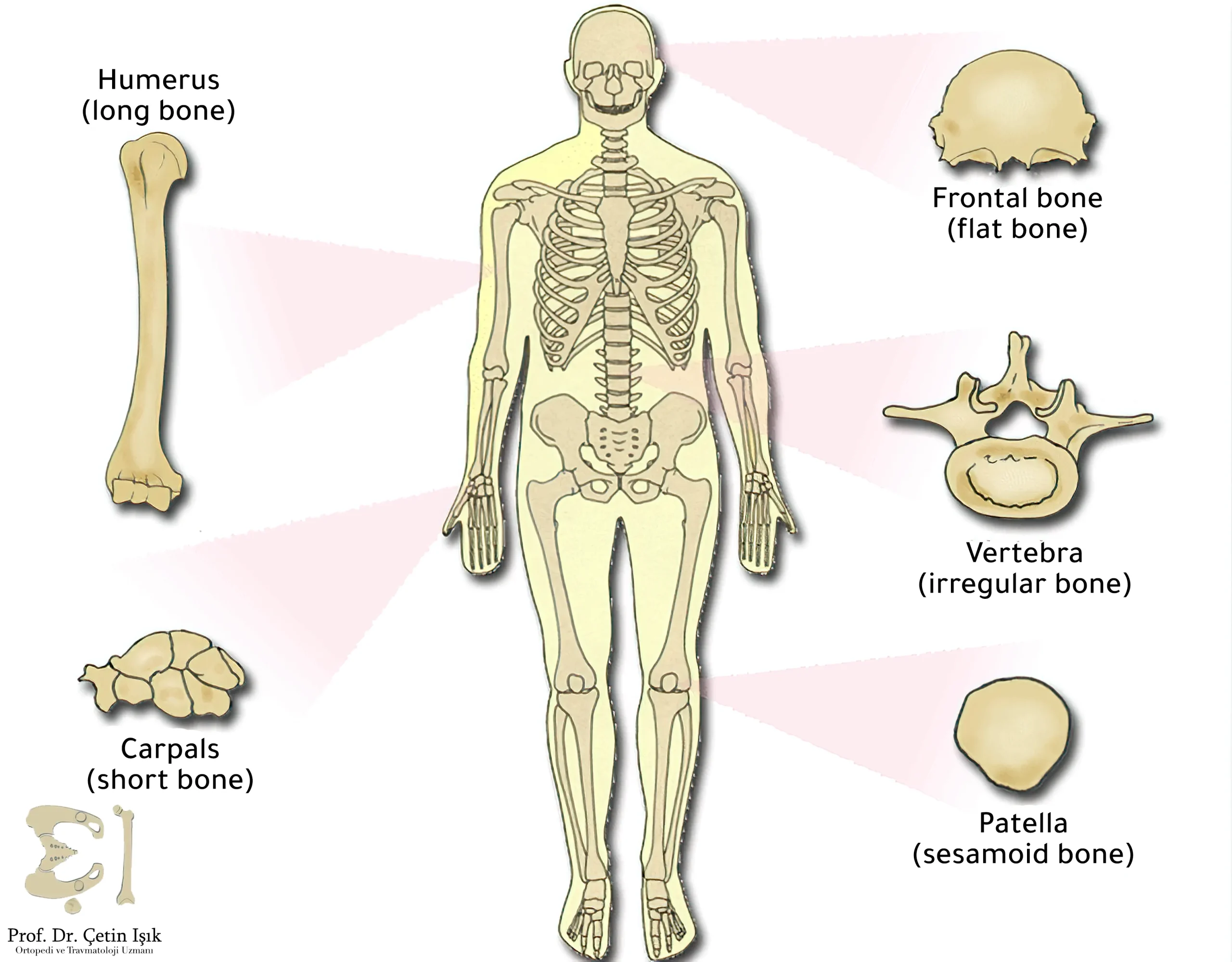
Bones are classified according to shape and structure into five groups: flat bones, short bones, long bones, irregular bones, and sesamoid bones.

Flat bones
It consists of two layers of dense bone called plates. The plates are separated by a layer of spongy bone tissue called the dipole. These bones are of comprehensive extension.
Examples: the walls of the cranial cavity (especially the occipital bone, the parietal bone, the frontal bone, the nasal bone, the lacrimal bone, and the vomer bone) - the scapula - the sternum.
Short bones
Bone core consists of spongy bone surrounded by dense bone. It has a cubic or square shape.
For example: the carpal bones of the hand (scaphoid bone - lunate bone - coccyx bone - trapezoid bone - triangular bone - large bone - canine bone) and the bones of the foot (calcaneus bone - navicular bone - talus bone or ankle bone - middle cuneiform bone - bone medial cuneiform).
Long bones
It consists of the bone body, a dense bone with a thin portion of cancellous bone on the inner surface around the bone marrow cavity, and epiphysis, which consists of spongy bone covered with a thin layer of dense bone.
Examples: the femur (the longest bone in the body) - fibula - tibia - metatarsal bones - humerus - radius - ulna.
These bones are characterized by several characteristics, the most important of which are:
- One of its diameters is larger than the other
- Its longitudinal bony axis is the largest
- It is distinguished by the presence of a middle part, which is the bone body
- The body of the bone ends with two ends, the epiphyses
- It contains a cavity inside the bone body that holds the bone marrow (for example, the femur and humerus)
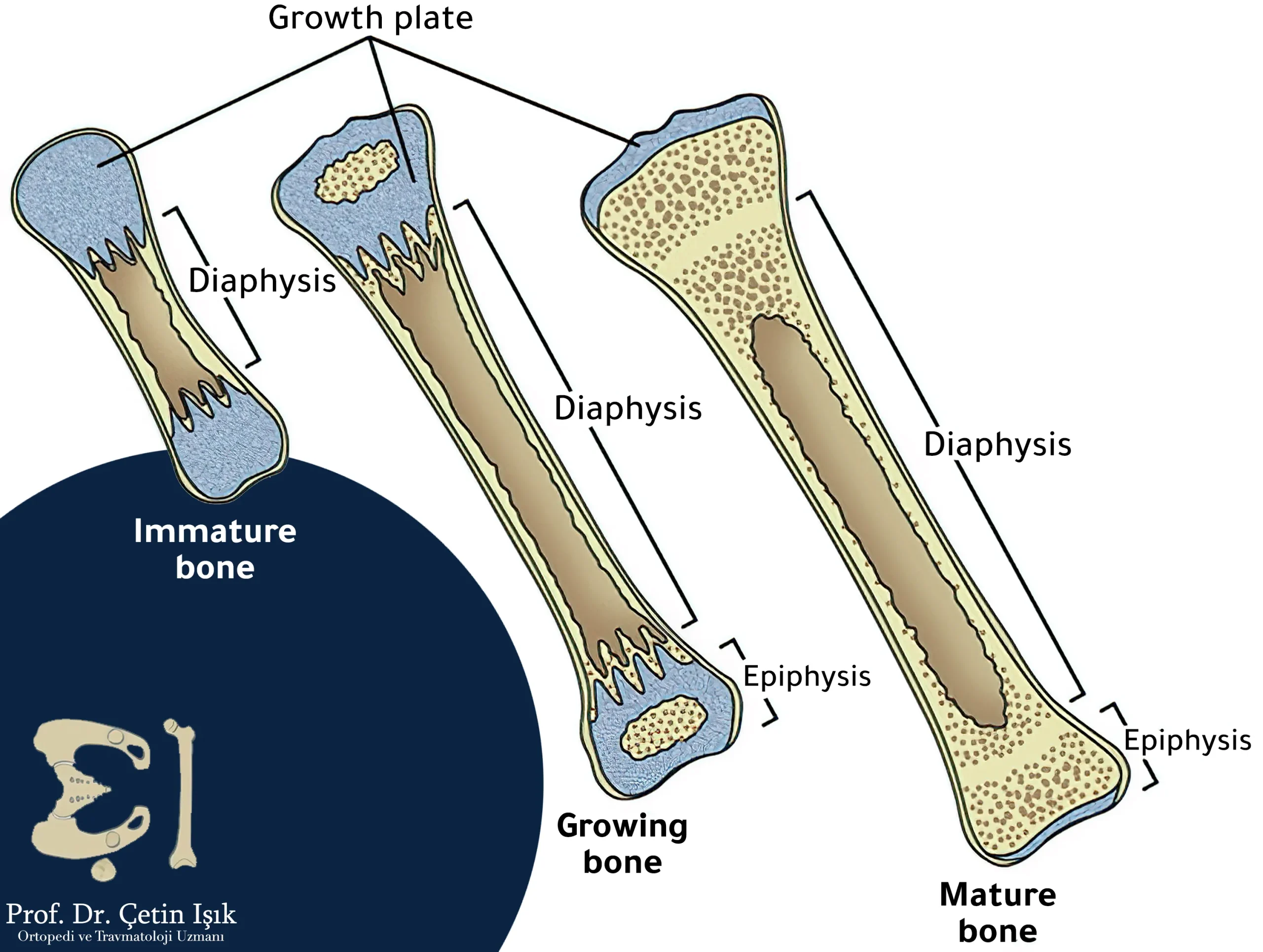
The long bone consists of:
- Epiphysis: The upper and lower end of a bone.
- Physis: It is located between the epiphysis and metaphysis.
- Metaphysis: A region connecting the growth cartilage (or epiphysis) and the body of the bone. The structure of this region is usually spongy.
- Diaphysis: Cortical bone containing bone marrow.
Irregular bones
Bone is classified under this type when it does not belong to any previous classes and does not have a definite bony shape.
These bones include, for example, the mandible, the vertebrae of the spine, the sphenoid bone, the pubis, the ilium, and the ischium.
Sesamoid bones
A group of small bones that take a round shape like a sesame seed, as its name suggests. Such bones are formed near the tendons (a particular tissue that connects the muscle to the bone).
The function of the sesamoid bones is manifested in relieving pressure on the joints and maintaining the tendon by strengthening it to overcome the compressive forces to which it is exposed.
These bones are located on the front surface of the joints, and the patella in the knee joint is the most prominent sesamoid bone in the body.
The bony cavities formed by bones
The bone cavity is a hole filled with air or fluid (joint fluid). The bony cavities in the human body are divided into:
- articular bony cavities:
The most common and most important, e.g., the glenoid cavity in the upper limb.
- Non-articular bony cavities:
It is more numerous than the first, including the air cavities, which are spaces between bones filled with air (such as the maxillary sinus and the tympanic cavity in the middle ear).
- Containing bony cavities:
It contains a group of organs, e.g., the cerebellar fossa and the orbital bone.
- Cavities for muscule tendon attachments;
These cavities give muscles a more significant fulcrum, for example, the pterygoid fossa, which includes muscles responsible for chewing and swallowing movements.
In conclusion, knowing the types of bone is essential to understanding the injuries that occur to them and knowing the best way to treat each bone according to its type.
Sources:
- Visible body
- Junqueira's Basic Histology – Bone tissue/chapter 8 – Types of bone page 143
- Gray's anatomy
Common questions
The bone is short when its length is approximately equal to its width, and short bones are considered one of the five types of bone, and an example is the wrist bones.
Bone growth begins with its transformation from its cartilaginous form to the stage of solid bone, so children often have a more significant number of cartilaginous bones that may exceed 300 bones, but the number of bones in the skeleton is approximately 206, according to what was mentioned in academic anatomy books.
The long bones are among the most important bones of the body. The femur is the longest bone in the human skeleton, as it constitutes nearly a quarter of its length and is considered the strongest and most solid bone in its body. It is found in the lower limb articulated with the pelvic girdle at the top and the fibula and tibia bones from the shin bones at the bottom.
The three bones (malleus, incus, and stapes), known as the auditory ossicles in the middle ear, are the smallest in the body, the smallest of which is the staps. The auditory ossicles are the first bones to ossify during fetal development. They are connected and connect the tympanic membrane to the inner ear.
The lacrimal bone belongs to the category of flat bones and is considered the most fragile and vulnerable human bone. It is located in the orbital cavity and has two surfaces, one facing the eye and the other facing the nose.


