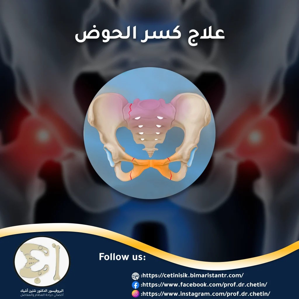
The treatment of a pelvic fracture may be emergency due to the large number of nearby vessels in addition to being a strong bone ring, so fractures of these bones often require strong trauma and are profuse bleeding.
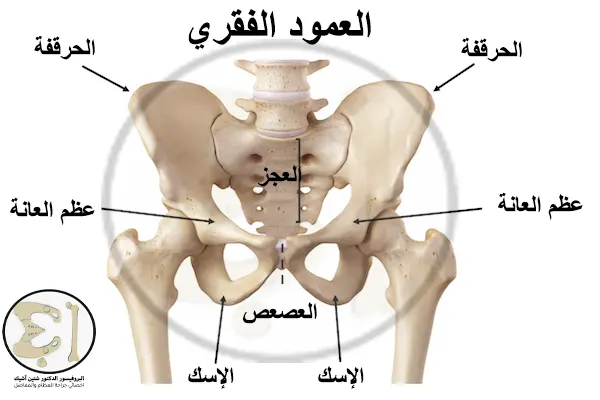
Anatomical profile of the pelvic bones
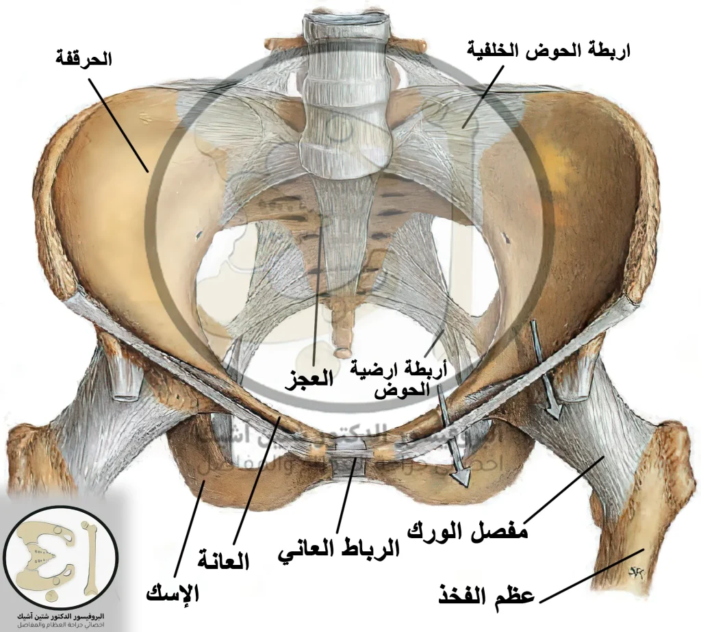
The ring of the pelvic bones consists mainly of three bones: the sacrum in the back and the two hip bones in front (also called the innominate bones).
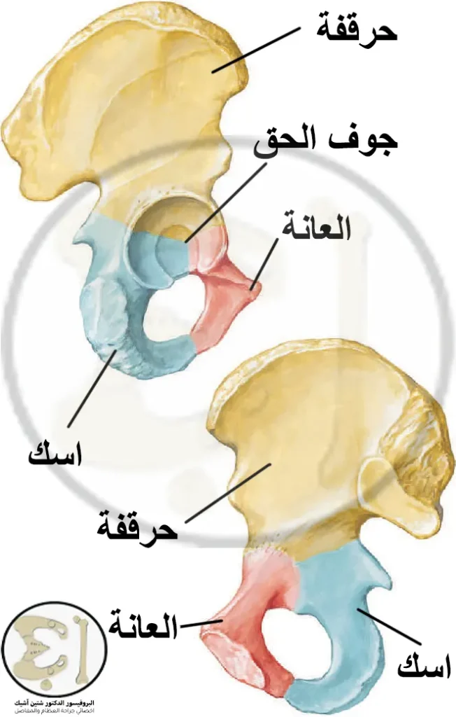
The sacrum consists of the body of the sacrum, the two wings of the sacrum, and the sacral promontory, and each hip bone is composed of three fused bones: the iliac, ischium, and pubis.
The pelvis is fixed by strong ligaments and is used in the classification of fractures, namely:
- Anterior ligaments: These include the pubic ligament
- The pelvic floor ligaments: They are two ligaments, the sacroiliac ligament, and the sacrotubercular ligament
- The posterior ligaments are the strongest ligaments in the human body. When a fracture occurs, the posterior ligaments are torn apart, which is evidence of the strong force of the shock leading to this fracture.
Classification of pelvic fractures
Pelvic fractures fall into several categories, which we will talk about:
Pelvic fractures by number of fractures
The fracture here is divided into a stable fracture and an unstable fracture
- stable fracture: There is a single pelvic ring fracture, and the two pieces of bone are attached.
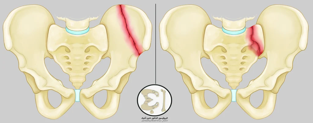
- Unstable fracture: There is more than one fracture in the pelvic ring and the bones are out of position.
Pelvic fractures according to the integrity of the skin
Here the fraction is divided into closed and open, in open fracture The bone protrudes from the skin and is more dangerous for the possibility of infection closed fracture The skin remains intact
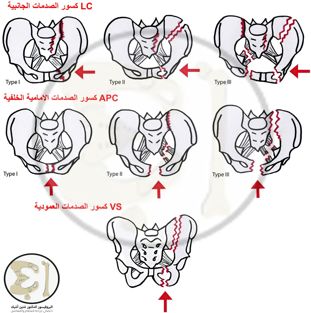
Fractures according to the direction of the trauma or the so-called Young-Burgess classification
Front Rear Fractures APC: It is three degrees differentiated by the rupture of the pelvic ligaments.
- The first degree is a rupture of the pubic ligament
- The second degree is a rupture of the pubic ligament and ligaments of the pelvic floor
- The third degree is a rupture of the pubic ligament, pelvic floor ligaments, and posterior pelvic ligaments
In third-degree fractures, the mortality rate is high, a lot of blood is lost, a significant need for a blood transfusion for the patient, and severe damage to the pelvic organs.
Side impact fractures LC: The occurrence of fractures in these shocks is more common than in anteroposterior shocks, and they are also of three degrees:
- The first degree is a fracture of the sacrum, with a fracture of one of the pubic or ischial branches on the same affected side.
- The second degree is a fracture of the iliac in addition to a fracture of one of the two branches of the pubic or ischium on the same affected side, and the shock here has a slightly forward direction.
- The third degree is the result of extreme lateral trauma; the fracture here is called the Windswept pelvis, and it manifests as a first or second-degree lateral fracture on the same side of the injury with a fracture on the opposite side to the side of the injury similar to an APC fracture
Vertical Traumatic Fractures VS: These fractures occur as a result of injuries to the side of the axis of the body, i.e. when falling from a height or in car accidents (for the driver), where the full shock load is on one foot, which leads to a fracture in one end of the pelvis, where the iliac crest rises above the normal level with a tear in the All ligaments of the pelvis.
Causes of pelvic fracture
The causes of pelvic fractures are divided into two categories:
Severe traumatic fractures: These injuries include falls from heights and car and motorcycle accidents, and they are often life-threatening fractures that need quick aid and are accompanied by bruises and fractures in the rest of the body with a lot of blood loss. The patient enters a state of shock, and most of these fractures are unstable.
Mild trauma fractures: The cause of these fractures is osteoporosis, especially in the elderly, because the weakness of the bones makes them vulnerable to fracture with less force, as an average fall in the bathroom or from the stairs may cause a fracture in the pelvic or hip bones, these fractures are usually of the stable type.
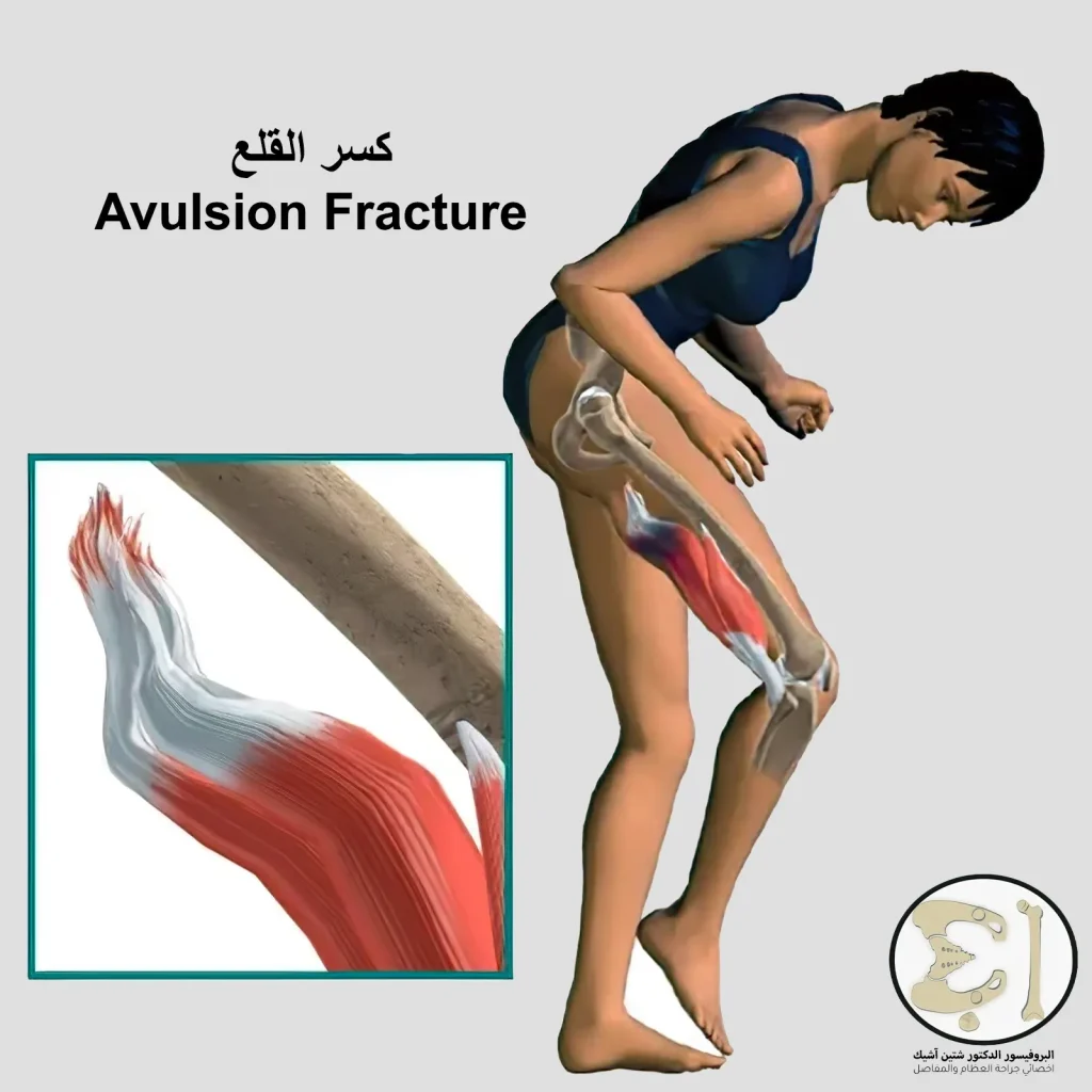
Avulsion FractureExtraction fractures occur in young athletes in the growth stage due to the large number of strenuous exercises that overload the great adductor muscle; thus, a piece of the ischial bone is removed from its place.
Types of pelvic and hip fracture
We must differentiate between two terms, pelvic fracture and hip fracture because pelvic fractures are in the bones that make up it (hip bones and sacrum).
hip fractures
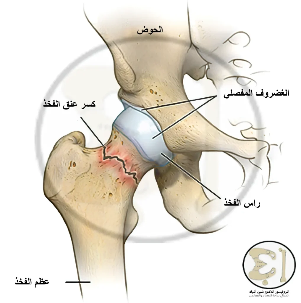
femoral neck fracture: It is a common fracture in the elderly, especially women over the age of 60, and is most often due to osteoporosis in old age, and it occurs as a result of a typical fall on the hip area. And we see that the injured leg is shorter than the other in these fractures (i.e., the two pieces of the femur that have been broken overlap).
femoral head fracture: It is a crack or fracture of the femoral head due to solid trauma such as traffic accidents, or it may occur if the bones are fragile due to repeated daily efforts such as walking a lot due to the constant pressure on the hip joint.
Trochanteric fracture of the thigh: The trochanter is located below the neck of the femur, and trochanter fractures occur due to a direct blow to the hip (the site of the trochanter) from a fall.
pelvic fractures
iliac bone fracture: An iliac fracture is characterized by swelling and pain in the groin area and the inability to put weight on the affected limb.
Pubic and ischial fractures: These fractures occur due to traffic accidents or falling from a high height, but, as we mentioned previously, extraction fractures may occur due to a sudden severe muscle contraction.
Acetabulum fracture: The acetabulum is an area of the hip bone in which the three bones (iliac, pubis, and ischium) come together to form a hole in which the femoral head resides, thus creating the hip joint. Fracture of the acetabulum is rare and occurs due to severe trauma.
Pelvic fracture symptoms
- Symptoms of a pelvic fracture vary according to the severity of the injury. Still, the main symptom is pain in the area of the fracture, which worsens with movement or placing weight on the pelvis (standing, for example).
- The fracture may be simple, so the patient feels discomfort when standing or walking (but he can stand or walk) with a little pain.
- Or the fracture may be severe, causing constant pain that prevents him from moving and does not ease until the patient places his foot in a position that comforts him (such as bending the knee). The goal of this position is to reduce weight from the pelvis or try to return the bones to their place.
- Symptoms may be swelling, puffiness, and bruising at the site of the broken pelvic bones.
Signs of a Pelvic Fracture Treatment
These are things that an orthopedic doctor or paramedics should note for rapid evaluation and treatment.
When a pelvic bone fracture occurs, it will not only affect the broken bone and the pain caused by it but will affect all nearby structures.
The most important and dangerous adjacent to the pelvic bones is large blood vessels These are the branches of the internal iliac artery and the external iliac artery. A fracture in the pelvis will lead to wounding these vessels, causing dangerous internal bleeding that may reach up to 4 liters (the volume of whole human blood is between 5.5 and 6 liters), and the medical team must correct this bleeding immediately.
This profuse bleeding will cause the patient to go into hypovolemic shock. We will see an increase in heart and respiratory rates, a decrease in pressure, pallor, and coldness in the extremities, and changes in the patient's consciousness. This bleeding is remedied by closing the bleeding vessel or tamping the bleeding area (putting gauze and pressure) in addition to an infusion of serums until the blood is secured.
One of the important neighborhoods is also nervesInjury to the nerves will lead to bad outcomes for the patient if he survives pelvic fractures.
Damage to the nerves -caused by a pelvic fracture- that are responsible for sensation or movement can lead to the inability to move the ankle or foot, loss of sensation in the sole, and in some cases, paralysis.
The nerves responsible for the genital area may be affected, leading to erectile dysfunction in men and impotence, while in women, it may lead to dyspareunia (pain during intercourse).
Pelvic fractures often damage the adjacent pelvic viscera. One of the most common male injuries is a urethral interruption. When the urethra is interrupted, we see a point of blood on the meatus at the head of the penis, and by rectal touch, we find the bladder high with a loose bloody mass below it. When the urethra is interrupted in females, we cannot install a Foley catheter (urinary catheter).
The rectum may also be affected, and here we notice the presence of blood in the anus, and we may also find blood in the vagina when it is injured.
In the future, pelvic fractures in women will increase the need for cesarean sections.
radiographic examination of pelvic fracture
The most crucial radiographic examination in emergency pelvic fractures is the emergency echogram, or the so-called FAST, which investigates the presence of blood pools indicating the presence of internal bleeding and directs us to treat the most severe cases first.
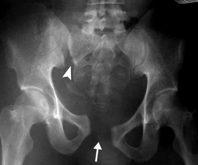
A pelvic X-ray is necessary to determine the classification of pelvic fractures and guide us in treatment.
Computed tomography (CT) scan is necessary to identify damage to adjacent tissues and blood vessels (when contrast media is injected).
Methods of treating a pelvic fracture
Treatment for a pelvic fracture varies according to the severity of the fracture, and treatment is divided into non-surgical treatment and surgical treatment.
Non-surgical treatment of pelvic fracture
Stable pelvic fractures are treated with non-surgical treatment, where the treatment depends on painkillers and reducing the burden on the broken bones until they heal, which is done by using crutches and wheelchairs, that is, without resorting to surgery.
We do not resort to surgical treatment here because the broken bone is not separated, and the fracture is single. There is no need for surgical intervention because it is possible to heal the fractured bone by itself.
The patient is also given anticoagulants and blood thinners to prevent clots from forming and releasing to the heart, or what is known as deep venous thrombosis (DVT).
Avoiding non-steroidal anti-inflammatory drugs (NSAIDs) is recommended, as they relieve pain but suppress the inflammatory process necessary for bone healing.
Despite the need for rest and not placing weights on the bone, a little movement is very necessary to prevent blood clotting as a result of stagnation and lack of movement. Movement is also necessary to prevent cases of muscle atrophy As a result of not moving it.
The latest non-surgical treatment methods for pelvic fracture
One of the most recent non-surgical methods of treating pelvic fracture is: Bone stimulation with low-intensity electricity or ultrasound.
This is done by applying daily low-intensity electrical pulses to the skin over the area of the broken bone. These pulses stimulate the bone cells to produce bone growth and healing components.
The patient may be given portable medical devices used to transmit electrical impulses for daily use for a period specified by the doctor.
For ultrasound, we apply a gel to the skin over the fracture area and place the ultrasound probe, which sends waves that accelerate the absorption of calcium and stimulate the bone cells to secrete substances that accelerate the healing process of the bone.
Surgical treatment of pelvic fracture
Patients with unstable pelvic fractures are the ones who need surgical intervention to restore the bones to their natural places. Still, we must remember that we are in front of an emergency patient and that treating pelvic fractures is not one of our priorities when he comes to the ambulance.
When a patient with an unstable pelvic fracture comes first, the integrity of the airway must be checked and that he is breathing, otherwise his breathing must be secured immediately, and then the presence of effective bleeding and the patient's need for blood transfusion must be checked.
After the patient's condition stabilizes and reaches safety, we consider treating the pelvic fracture.
Surgical pelvic fracture treatment patterns
A pelvic fracture is treated surgically by:
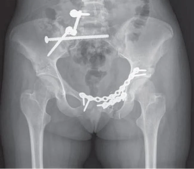
External repair in the treatment of pelvic fracture: It is the insertion of metal rods and screws into the broken bones by making small incisions in the skin and muscles to hold the fractured bones in place.
This method is used as a complete treatment procedure until the bones are completely healed or as a temporary measure because the patient cannot bear the surgery currently. Hence, we perform the external repair to treat pelvic fractures until the patient can tolerate open surgery.
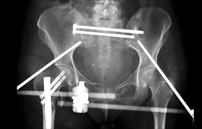
Internal repair in the treatment of pelvic fracture: Here, we perform the surgical opening to return the bones to their place and fix them with screws and metal plates.
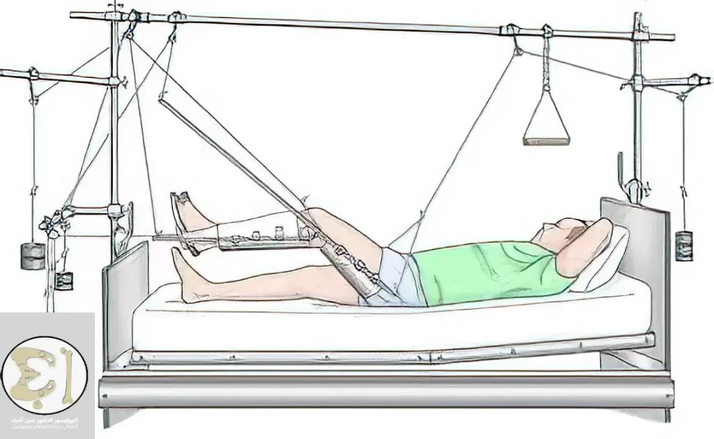
Skeletal Traction: Since the limb is shortened after a fracture of the hip or pelvis, we apply structural traction, which is two long metal rods on the ends of the leg with cross rods inserted into the femur or tibia, and we tighten the limb with some controlled weights and return the bones to their place.
Acetabulum fractures may be treated with skeletal traction alone, providing pain relief when the bones return to their place.
Complications of surgery in the treatment of pelvic fracture
Like other surgeries, pelvic fracture surgery has some complications, the most important of which are:
- Damage to blood vessels and nerves during surgery
- Problems closing the incision
- Blood clotting problems
- The worst is pulmonary embolism and pulmonary embolism
Postoperatively in the treatment of pelvic fracture
The doctor will prescribe pain relievers and blood thinners, and the doctor will encourage you to move without putting weight on the bones by using crutches.
He will also perform physical therapy sessions for rehabilitation, and the patient may be able to return to walking without crutches three months after surgery. Still, you may use crutches for a little longer.
In case you need any support for your health condition or want to inquire about the treatment of a pelvic fracture Contact with us.
We can answer everything you need or want to know about pelvic fracture treatment.
Sources:
Common questions
The recovery time of the pelvic fracture after treatment varies according to the severity of the fracture and the type of treatment provided. Still, the patient is expected to be able to walk three months after the surgical treatment of the pelvic fracture.
Pelvic fractures heal if appropriate treatment is provided according to the fracture type. Still, the risk lies in complications of the fracture, such as damage to the viscera and adjacent nerves, as well as the possibility of women needing a cesarean section in the future in the event of pregnancy and childbirth after pelvic fractures.
In most cases, pelvic fractures are dangerous because the formation of the pelvic bones and how they are placed gives them strength and durability. Therefore, a pelvic fracture requires a strong force, which occurs in car accidents and falls from high heights. Hence the danger of pelvic fractures will come from two factors:
First, the presence of large blood vessels adjacent to the pelvis will lead to severe internal bleeding in the event of a pelvic fracture.
Secondly, the presence of such severe injuries in the pelvis necessitates the presence of damage in other places, such as the head and chest, and bruises in these places may be fatal.
Putting weight on the broken pelvis will make it worse, so you will not be able to walk at first and will stay in bed for a certain period. Doctors will advise you to walk with crutches instead with some physical treatments to restore your muscle strength, and after the bone heals completely, you will start walking gradually.
Pelvic fractures begin to heal after about four to six weeks.
In some cases where the pelvic fracture is stable (a single fracture and the two bony pieces are not separated, that is, the bone is only cracked), the pelvic fracture can be treated by relying on pain relievers until it heals on its own.