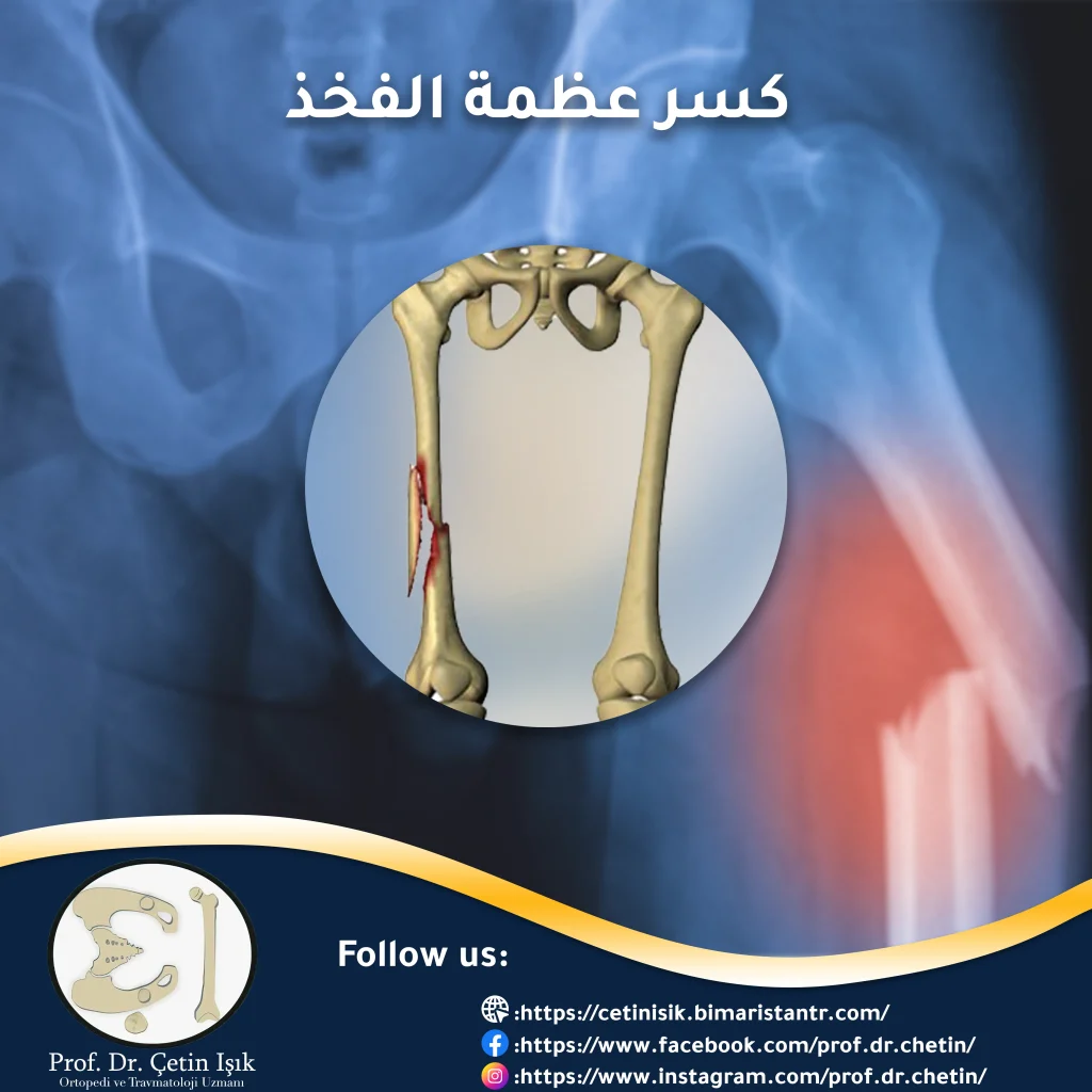
The femoral fracture is one of the serious fractures that require emergency treatment, given that the femur is the strongest bone in the body, so the fracture often occurs in it due to strong trauma.
A fracture of the femur poses a threat to human life, especially the elderly. You should not hesitate for a single moment in thinking of providing first aid to patients with femur fractures. Continue in this article to learn more about these fractures that carry great risks and serious complications.
Introduction to femur fracture
It is a separation of the bone tissue in different anatomical places of the thigh due to a very severe injury, which needs immediate attention.
The long femur is one of the longest and strongest bones of the lower extremity, which connects the hip joint at the top with the knee joint at the bottom, in addition to being the basis for many ligaments and tendons.
Measures and complications differ according to the fracture sites in the femur, and each fracture in the femur has a certain age, so it is important to know the classification of bone fractures in the femur.
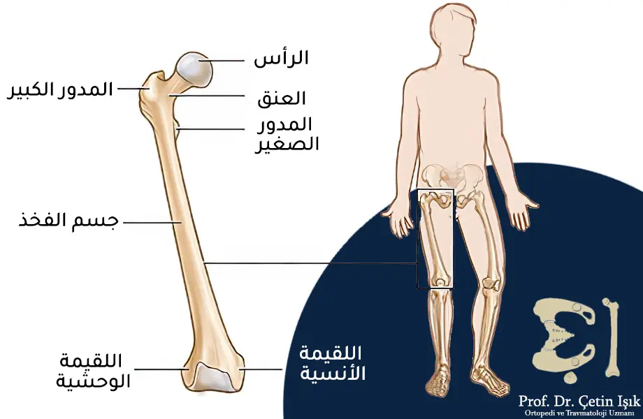
Location of hip fractures
femoral neck fracture Trochanteric fractures, in addition to the fracture of the femoral head, are classified under the classification of hip fracture or intra-articular fractures. As for condylar fractures (lateral and medial), they are classified with fractures of the knee joint. The body of the thigh is divided into three parts where the fracture occurs, including:
- one-third of the distal femur
- middle third
- proximal third
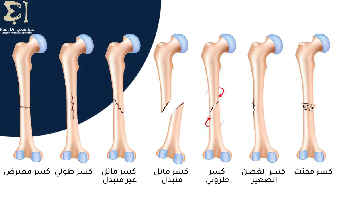
Classification of different types of femur fractures
The types of femur fractures are divided into:
- Transverse fracture: It is a straight horizontal line that divides the body of the thigh.
- Oblique fracture: an oblique fracture of the body of the femur.
- Comminuted fracture: The bone breaks down into small bony pieces.
- Spiral fracture: surrounds the body of the femur.
- Variable fracture: Occurs when both sides of the femur fracture separate from each other.
- Open fracture: It occurs when the muscles and tendons are damaged as a result of the bone breaking through the skin tissue.
- Small branch fracture: a slight crack in the bony edges, without breaking.
- Double fracture: Comprehensive fracture of two different positions in the body of the femur.
Causes of femur fracture
Femoral fractures occur most commonly in the elderly due to the fragility of the femur in the elderly or Osteoporosis They have it, but it can happen at any age. Most cases are due to traumatic causes, usually such as trauma or injuries in traffic accidents, such as car and motorcycle accidents, or falling from a great height. Also, gunshots cause such fractures in the bones.
Symptoms and signs of a femur fracture
When the femur is fractured, one or more of the following signs appear, which are evidence of the fracture:
- Severe pain (the most common symptom) and the inability to walk or put weight on the injured leg
- Swelling at or around the affected area
- Deformation of the affected limb
- Shortness of the affected limb compared to the healthy limb
- Thigh bone misalignment
- Hemorrhage
- Abnormal movement of the affected limb
Complications of a femur fracture
Complications of a femur fracture include:
- Heavy bleeding, especially in open and comminuted femur fractures, which can predispose to shock leading to death.
- Stiffness in the knee joint occurs if the patient is not rehabilitated early.
- Non-healing or its delay.
- Healing and defective healing.
- A child's growth may be affected if the epiphysis is affected.
- Dry necrosis in cases of fractures of the neck of the femur, which may be associated with a fracture of the femur, as may result from these accompanying fractures. degenerative disease in the right hollow.
- Articulated stiffness.
Diagnosing a femur fracture
Diagnosis involves several steps, including:
Clinical history and clinical examination
The injured person or one of the accompanying persons is asked about the details that led to the fracture of the thigh or the patient’s injury and how they were exposed to the accident, such as if he was exposed to a fall accident or cases of falling from a high height or a traffic accident. All this helps doctors to assess the extent of the injury. Accompanying disease or previous diseases, or if he is taking certain medications, then he moves to the clinical examination in which the therapists contemplate most areas of the body and examine the general health condition, and make sure that the rest of the bones are intact from fracture or dislocation, then focus on the thigh and leg in addition to the pelvis where they are examined Thigh muscle strength and range of motion.
Radiographic diagnosis
X-Ray and CT scans help diagnose a femur fracture and determine the type, location, and severity of the femur fracture. CT scans are more accurate and can show minor cracks that X-rays don't show.
Treatment of a femur fracture
Developing a treatment plan for a femur fracture depends on the location, type, and severity of the injuries occurring in the femur. Treatment is divided into:
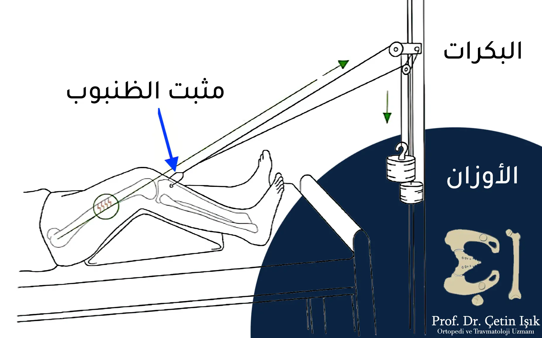
structural extension
A resistant stretching of the muscles that replace the fracture, applied by a skewer inserted through the femur and with local anesthesia, we resort to it in the event that surgery is not preferred, such as the difficulty of general anesthesia due to health problems, or in the case of severe comminuted fractures, or in the event of difficulty in stabilizing the bones.
Internal fixation
It uses metal anchors such as wires, skewers, plates, and screws, to stabilize the bones until healing occurs and the femur is completely repaired.
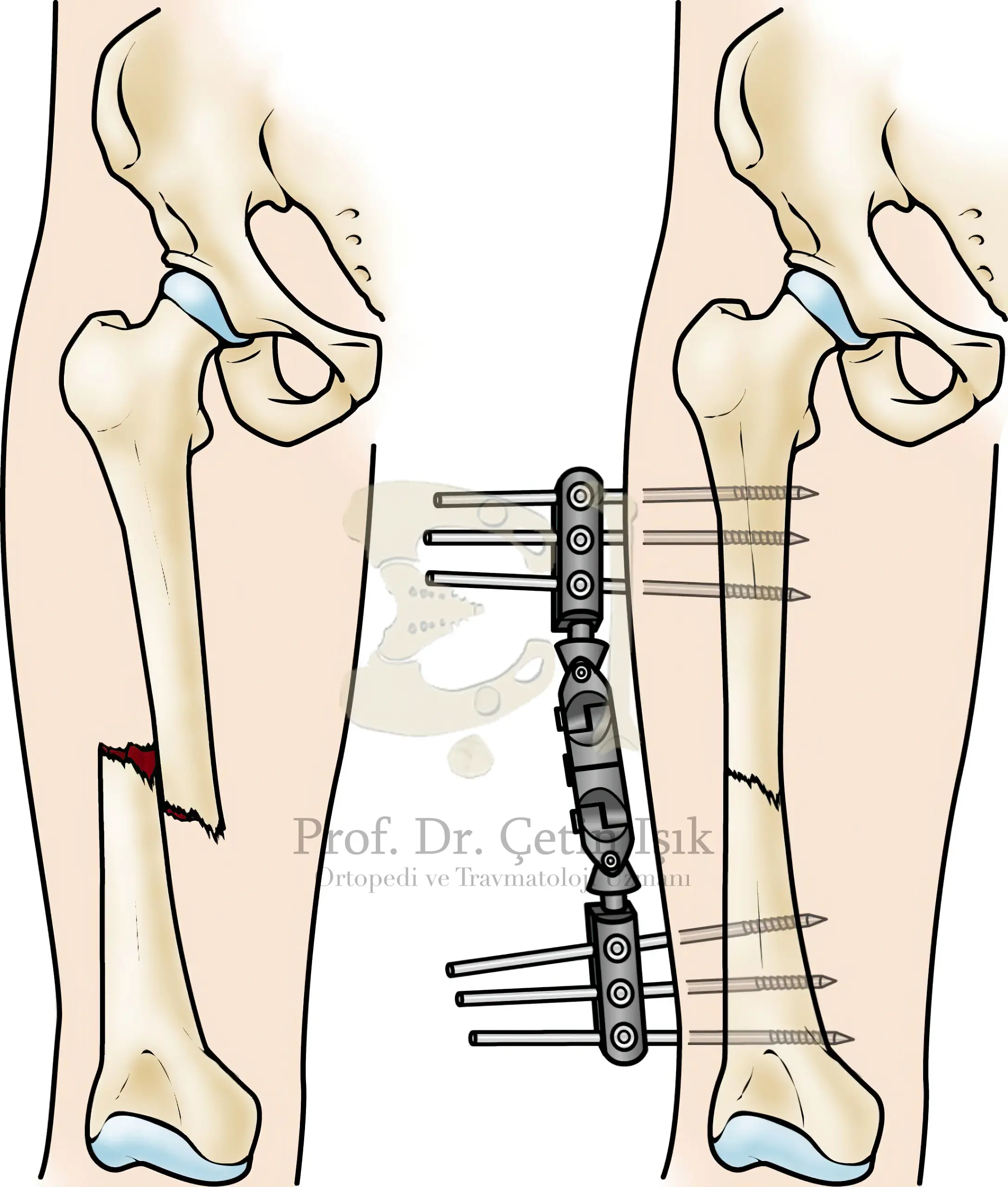
external install
An effective and quick way to repair bones, in which metal skewers are used, which we insert through the skin and muscles to reach the bone (above and below the fracture) without going through the focus of the fracture, thus fixing the fracture.
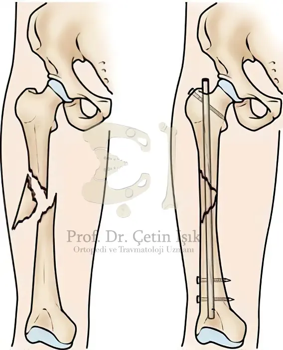
Intrinsic sphenoids (medullary screws)
We do not open the focus of the fracture, a channel is drilled in the knee or trochanter by radioscopy, then a skewer is inserted that fills the channel, then screws are installed in the thigh joint to fix it.
Physical rehabilitation
After surgery on a broken bone, physical rehabilitation doctors give exercises to help restore normal movement.
Other medical information
- The fracture in the upper part of the femur, in addition to closed femur fractures, is treated with surgical treatment.
- Conservative treatment is often preferred in femoral fractures in children, due to their faster recovery.
- Paracetamol and other painkillers can be given to control the pain of a broken bone.
- Treatment of a hip fracture, or treatment of a hip fracture, can be done bHip replacement surgery.
In conclusion, femoral fractures pose a threat to the life of the elderly. The measures and complications of the types of femoral fractures differ, so it is very important to diagnose the location and type of fracture to ensure the correct and appropriate treatment.
Sources:
Common questions
The fact that the thigh bone is the largest bone in the body makes this bone the most important bone in the body, in addition to the frequent occurrence of complications when a thigh fracture occurs, such as profuse bleeding and infection, which can lead to death if the patient’s condition worsens.
The duration of the healing of the femur bones depends on the location, severity and type of fracture, but the average recovery period is estimated from 16 to 18 weeks and may increase or decrease by a few days. Longer than usual due to the fragility of their bones and joints.
After performing the hip fracture operation or after removing the gypsum by the doctor’s decision, it is possible to walk with crutches and do special exercises to restore the muscles to their previous strength, and after one to two months the normal movement returns somewhat with following the doctors’ recommendations to avoid a new fracture.
In the event of an injury to the bone epiphysis and growth plate in children, the affected limb becomes shorter than the healthy limb.